Acute Haemorrhage
Summary
- Acute haemorrhage is the rapid loss of blood from the circulatory system due to vascular injury or underlying medical conditions
- Clinical presentation varies based on location and severity, ranging from localised pain to hypovolemic shock
- Imaging plays a crucial role in identifying the source and extent of bleeding, guiding treatment decisions
Pathophysiology
- Caused by disruption of vascular integrity due to:
- Trauma
- Aneurysm rupture
- Arteriovenous malformations
- Coagulopathies
- Iatrogenic injuries
- Results in:
- Decreased blood volume
- Reduced tissue perfusion
- Potential organ dysfunction
Demographics
- Incidence varies based on etiology:
- Trauma: Higher in young adults and males
- Spontaneous intracranial haemorrhage: Increases with age
- Gastrointestinal bleeding: More common in elderly populations
- Risk factors include:
- Hypertension
- Anticoagulant use
- Liver disease
- Genetic predisposition
Diagnosis
- Clinical presentation:
- External bleeding: Visible blood loss
- Internal bleeding: Pain, swelling, haemodynamic instability
- Laboratory tests:
- Complete blood count
- Coagulation profile
- Liver function tests
- Imaging studies:
- CT, MRI, ultrasound, angiography
Imaging
- Computed Tomography (CT):
- Rapid assessment of multiple body regions
- Contrast-enhanced CT for active bleeding detection
- CT angiography for vascular injuries
- Magnetic Resonance Imaging (MRI):
- Superior soft tissue contrast
- Useful for subacute and chronic haemorrhage
- Limited in acute settings due to longer acquisition times
- Ultrasound:
- FAST protocol in trauma settings
- Doppler studies for vascular assessment
- Angiography:
- Gold standard for detecting active bleeding
- Allows for simultaneous intervention
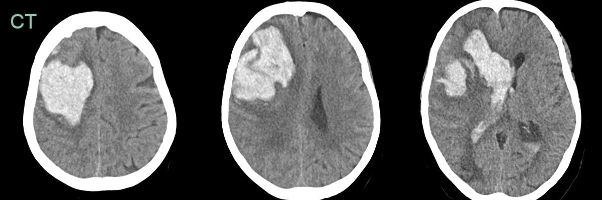
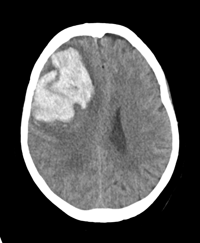
- A hyperdense haematoma, with a thin rim of surrounding oedema, in the right frontal lobe has discharged into the right lateral ventricle.
Treatment
- Initial management:
- Haemodynamic stabilization
- Blood product transfusion as needed
- Surgical intervention:
- Exploratory laparotomy/thoracotomy
- Neurosurgical procedures for intracranial haemorrhage
- Endovascular techniques:
- Embolization
- Stent placement
- Medical management:
- Reversal of anticoagulation
- Correction of coagulopathies
- Blood pressure control
Differential diagnosis
| Differential Diagnosis (and causes of haemorrhage) | Differentiating Feature |
|---|---|
| Haemorrhagic transformation of an infarct | Arterial territory diffusion restriction on MRI beyond the haematoma |
| Subdural Haemorrhage | Crescentic extra-axial collection crossing sutures |
| Epidural Haemorrhage | Biconvex extra-axial collection not crossing sutures |
| Tumour | Mass effect, surrounding oedema, enhancement with contrast |
| Contusion | History of trauma, coup-contrecoup pattern |
| Venous Sinus Thrombosis | Empty delta sign, cord sign on CT/MRI |
| Arteriovenous Malformation | Serpiginous flow voids, calcifications |
| Amyloid Angiopathy | Lobar microhaemorrhages, superficial/lobar distribution |
| Arteriolosclerosis | Basal ganglia small vessel disease and microhaemorrhages |
| Coagulopathy | Abnormal coagulation profile, multiple bleeds |