Balo's concentric sclerosis
Summary
- Rare demyelinating disorder characterised by concentric rings of alternating preserved and destroyed myelin
- Typically presents with acute or subacute neurological deficits
- MRI shows pathognomonic concentric ring lesions with alternating bands of T2 hyperintensity and isointensity
Pathophysiology
- Considered a variant of multiple sclerosis (MS)
- Exact etiology unknown, but proposed mechanisms include:
- Alternating bands of cytokine expression leading to concentric demyelination
- Hypoxia-induced tissue injury with preservation of small veins
- Lesions typically occur in white matter, but can also affect gray matter
Demographics
- Rare disease, with fewer than 100 cases reported in literature
- More common in young adults (20-40 years old)
- Higher prevalence in East Asian populations
- No clear gender predilection
Diagnosis
- Clinical presentation:
- Acute or subacute onset of neurological deficits
- Symptoms depend on lesion location, may include:
- Motor weakness
- Sensory disturbances
- Visual impairment
- Cognitive changes
- Cerebrospinal fluid analysis:
- May show elevated protein and oligoclonal bands (similar to MS)
- Diagnosis primarily based on characteristic MRI findings
Imaging
- MRI is the imaging modality of choice
- Characteristic findings:
- Concentric rings of alternating signal intensity on T2-weighted images
- T1-weighted images: Hypointense rings
- T2-weighted images: Alternating hyperintense and isointense bands
- FLAIR: Similar appearance to T2-weighted images
- Contrast enhancement: Variable, may show incomplete ring enhancement
- Diffusion-weighted imaging (DWI):
- May show restricted diffusion in acute lesions
- MR spectroscopy:
- Decreased N-acetylaspartate (NAA) and increased choline and lactate peaks
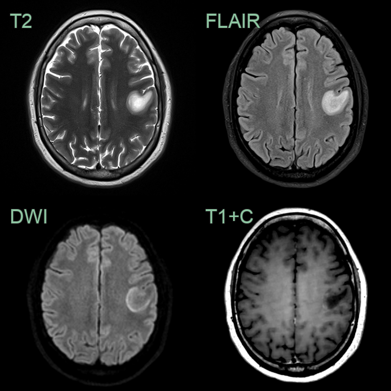
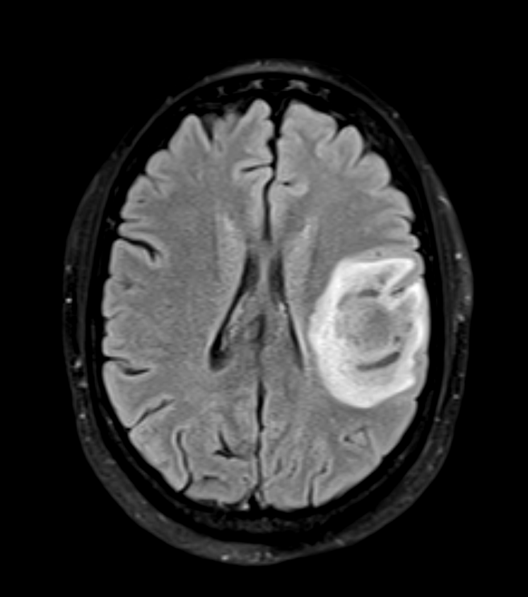
- A 40-year-old patient presented with right-sided sensory disturbance. Initial MRI showed a T2-hyperintense lesion in the left precentral gyrus with a rim of diffusion restriction and subtle enhancement.
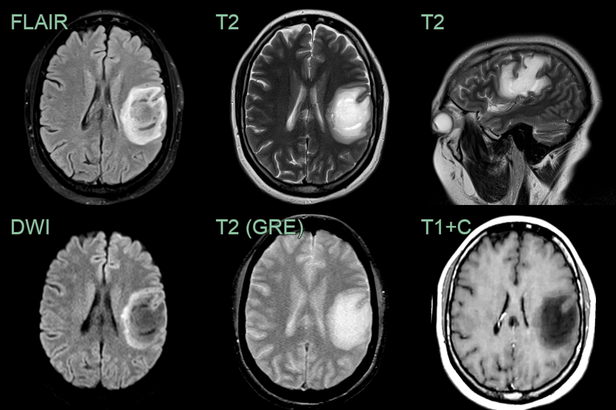
- Despite steroid therapy, an MRI one month later showed that the lesion has significantly enlarged although the enhancement resolved.
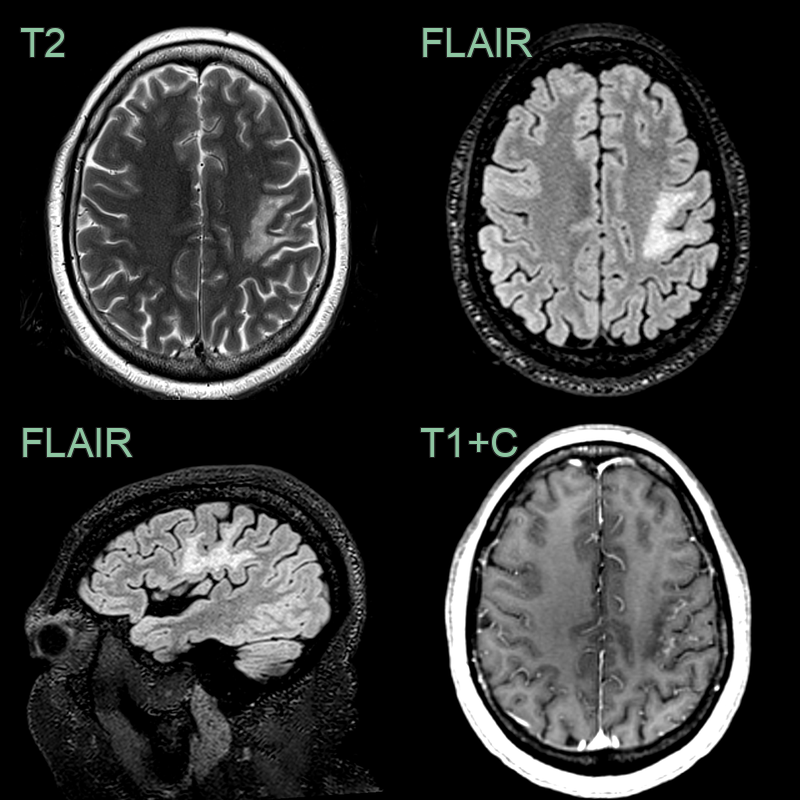
- After two months, following plasma exchange, the lesion had decreased in size although punctate enhancement had developed.
Treatment
- No standardized treatment protocol due to rarity of the disease
- Management approaches similar to those used for acute MS relapses:
- High-dose intravenous corticosteroids
- Plasma exchange for steroid-refractory cases
- Immunomodulatory therapies:
- Interferon-beta, glatiramer acetate, or other MS disease-modifying drugs may be considered for long-term management
- Supportive care and rehabilitation for neurological deficits
- Prognosis:
- Variable, ranging from complete recovery to severe disability
- Some cases may progress to MS or show recurrent Balo-like lesions
Differential diagnosis
| Differential Diagnosis | Differentiating Feature |
|---|---|
| Multiple Sclerosis | Lacks concentric ring pattern on MRI; typical ovoid periventricular plaques |
| Acute Disseminated Encephalomyelitis | Diffuse, bilateral white matter lesions without concentric ring pattern; basal ganglia involvement common |
| Tumefactive Demyelinating Lesions | Large, solitary lesions without concentric ring pattern; incomplete ring enhancement open towards cortex |
| Neuromyelitis Optica Spectrum Disorder (NMSOD) | Predominant optic nerve and long spinal cord lesions; area postrema involvement |
| Primary CNS Lymphoma | Homogeneous enhancement on MRI; restricted diffusion; periventricular location; hyperdense on non-contrast CT |
| Progressive Multifocal Leukoencephalopathy | Irregular non-enhancing lesions with restricted diffusion; subcortical U-fibre involvement; no ring pattern |
| Cerebral Infarction | Follows vascular territory; wedge-shaped; DWI restriction in acute phase; no ring pattern |
| Cerebral metastasis | Multiple lesions at grey-white junction; ring or nodular enhancement without concentric pattern |
| Cerebral Abscess | Ring-enhancing lesion with restricted diffusion and surrounding oedema |