Capillary Telangiectasia
Summary
- Benign vascular malformation composed of dilated capillaries
- Typically asymptomatic and found incidentally on imaging
- Most commonly located in the pons, but can occur throughout the brain
Pathophysiology
- Characterised by dilated capillary-sized vessels without intervening brain parenchyma
- Lack of smooth muscle and elastic fibres in vessel walls
- No evidence of cellular proliferation or neoplasia
- May be associated with developmental venous anomalies (DVAs) in some cases
Demographics
- Prevalence: 0.4-2% in autopsy series
- No significant gender predilection
- Can occur at any age, but most commonly diagnosed in adults
Diagnosis
- Usually asymptomatic and discovered incidentally on imaging
- Rarely associated with minor neurological symptoms:
- Headaches
- Dizziness
- Focal neurological deficits (uncommon)
- Differential diagnosis includes:
- Low-grade gliomas
- Demyelinating lesions
- Small infarcts
Imaging
- MRI is the modality of choice for diagnosis
- T1-weighted imaging:
- Isointense or slightly hypointense to brain parenchyma
- T2-weighted imaging:
- Mildly hyperintense
- T2*-weighted imaging:
- May show "brush-like" appearance due to dilated vessels
- Post-contrast T1-weighted imaging:
- Mild, homogeneous enhancement
- "Brush-like" or "stippled" enhancement pattern
- No mass effect or surrounding oedema
- No restricted diffusion on DWI
- DSC perfusion:
- No significant increase in relative cerebral blood volume (rCBV)
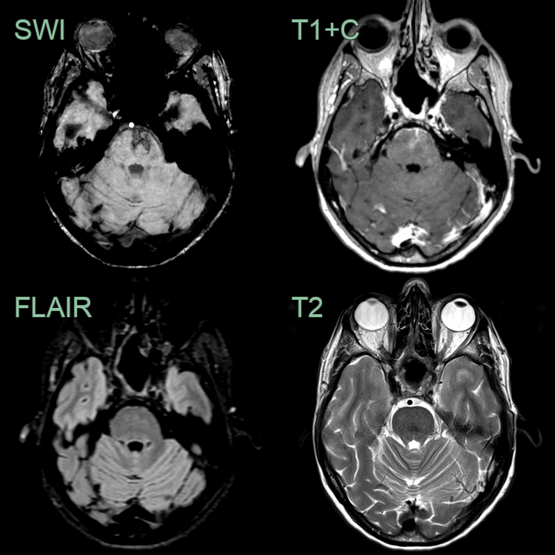
- An incidental lesion shows reticular susecptibility artefact, hazy enhancement and is invisible on T2-weighted imaging.
Treatment
- No treatment required for asymptomatic lesions
- Regular follow-up imaging may be recommended to ensure stability
- Symptomatic cases (rare):
- Conservative management of symptoms
- Surgical intervention generally not indicated due to benign nature and risk of complications
- Patient education and reassurance about the benign nature of the lesion
Differential diagnosis
| Differential Diagnosis | Distinguishing Feature |
|---|---|
| Cavernous Malformation | Lacks haemosiderin rim on MRI; no mass effect |
| Developmental Venous Anomaly | Has characteristic "caput medusae" appearance on contrast-enhanced imaging |
| Arteriovenous Malformation | Lacks arteriovenous shunting on angiography |
| Cerebral Metastasis | Lacks surrounding oedema and mass effect; no enhancement on contrast MRI |
| Glioma | No mass effect or surrounding oedema; lacks contrast enhancement |
| Multiple Sclerosis Plaque | Lacks periventricular predilection; no enhancement on contrast MRI |
| Acute Small Infarct | No restricted diffusion on DWI; lacks evolution over time |
| Petechial Haemorrhage | No blooming artefact on susceptibility-weighted imaging |
| Hypertensive Microbleeds | Lacks preferential distribution in basal ganglia, thalamus, or cerebellum |
| Radiation-Induced Telangiectasia | Located within prior radiation field; may have associated white matter signal change and radiation necrosis |