Central Vein Sign
Summary
- Radiological finding in multiple sclerosis (MS) characterised by a central vein within white matter lesions
- Helps differentiate MS from other white matter diseases, particularly small vessel disease
- Visible on susceptibility-weighted imaging (SWI) and T2*-weighted MRI sequences
Pathophysiology
- MS lesions typically develop around small veins in the white matter
- Inflammation and demyelination occur around these central veins
- The central vein remains visible within the lesion due to:
- Increased deoxyhaemoglobin content
- Magnetic susceptibility differences between the vein and surrounding tissue
Demographics
- Most commonly observed in patients with MS
- Can be seen in all subtypes of MS:
- Relapsing-remitting MS
- Secondary progressive MS
- Primary progressive MS
- Less frequently observed in other white matter diseases
Diagnosis
- Central vein sign is a supportive feature in MS diagnosis
- Criteria for positive central vein sign:
- Vein visible in the centre of the lesion
- Vein runs partially or entirely through the lesion
- Vein visible in at least two perpendicular planes
- Proposed diagnostic threshold:
-
40% of white matter lesions should demonstrate the central vein sign for MS diagnosis
-
Imaging
- Best visualised on:
- Susceptibility-weighted imaging (SWI)
- T2*-weighted sequences
- 3T MRI provides better visualisation than 1.5T
- FLAIR* imaging (combination of FLAIR and T2*) can improve detection
- Imaging parameters:
- High spatial resolution
- Thin slices (≤3mm)
- Minimal slice gap
- Post-processing techniques:
- Minimum intensity projection (mIP)
- Segmented-EPI for improved resolution
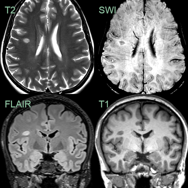
- A patient with a diagnosis of MS had an ovoid lesion in the right frontal subcortical white matter.
- SWI showed that the lesion was centred on a medullary vein.
Treatment
- Central vein sign does not directly influence treatment decisions
- Aids in early and accurate diagnosis of MS, which can lead to:
- Earlier initiation of disease-modifying therapies
- Improved long-term outcomes
- Potential applications in clinical trials:
- Patient selection
- Monitoring treatment response
- Future developments may include:
- Automated detection algorithms
- Standardisation of imaging protocols for widespread clinical use
Differential diagnosis
| Differential diagnosis | Differentiating feature |
|---|---|
| Multiple Sclerosis | Central vein sign present in >40% of lesions |
| Small Vessel Disease | Lesions typically do not have central vein |
| Neuromyelitis Optica | Lesions tend to be larger and follow different distribution |
| Acute Disseminated Encephalomyelitis | Lesions are typically larger and more confluent |
| Cerebral Vasculitis | Lesions often follow vascular territories |
| Susac Syndrome | Characteristic involvement of corpus callosum "snowball" lesions |
| Migraine with Aura | No visible lesions on MRI |
| CADASIL | Characteristic involvement of anterior temporal lobes and external capsule |
| Lyme Disease | Lesions are typically non-specific and may resolve with treatment |
| Progressive Multifocal Leukoencephalopathy | Lesions are typically larger and in subcortical white matter |