CTA Dot Sign
Summary
- The CTA dot sign refers to a focal hyperdense dot within a cerebral artery on CT angiography
- It indicates the presence of an intraluminal thrombus in acute ischaemic stroke
- The sign is highly specific for vessel occlusion and aids in early diagnosis and treatment planning
Pathophysiology
- Represents a cross-sectional view of an intraluminal thrombus within a cerebral artery
- Typically seen in M1 segment of the middle cerebral artery (MCA) or basilar artery
- Composed of red blood cells, platelets, and fibrin, causing local hyperdensity on CT
Demographics
- Primarily observed in patients with acute ischaemic stroke
- More common in older adults due to higher stroke incidence
- No significant gender predilection reported
Diagnosis
- Clinical presentation:
- Sudden onset of neurological deficits
- Symptoms vary based on the affected vascular territory
- CT angiography (CTA) is the primary diagnostic modality
- Differential diagnosis:
- Calcified atherosclerotic plaque
- Contrast material in small perforating arteries
Imaging
- CT angiography findings:
- Hyperdense dot within the lumen of an affected artery
- Most commonly seen in M1 segment of MCA
- Size typically 1-2 mm in diameter
- Associated findings:
- Vessel cutoff sign distal to the dot
- Reduced contrast opacification in distal branches
- Sensitivity and specificity:
- High specificity (>95%) for vessel occlusion
- Moderate sensitivity (50-70%) due to small size and partial volume effects
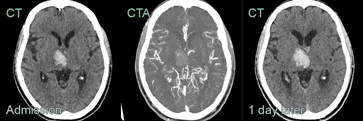
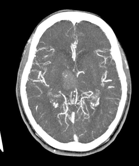
- A 60-year-old patient presented with left sided weakness and parasthesia.
- Imaging showed a focus of enhancement within a right thalamic haematoma.
- A follow-up CT showed enlargement of the haematoma.
Treatment
- Rapid identification of CTA dot sign guides treatment decisions
- Intravenous thrombolysis with recombinant tissue plasminogen activator (rtPA) within 4.5 hours of symptom onset
- Mechanical thrombectomy for large vessel occlusions:
- Considered up to 24 hours in selected patients
- Particularly beneficial for M1 MCA occlusions
- Antiplatelet therapy and management of risk factors for secondary prevention
Differential diagnosis
| Differential Diagnosis | Differentiating Feature |
|---|---|
| Cortical vein thrombosis | CTA dot sign is located in subarachnoid space, not within cortical vein |
| Aneurysm | CTA dot sign is smaller and lacks the typical saccular shape of aneurysms |
| Arteriovenous malformation | CTA dot sign lacks the characteristic tangle of abnormal blood vessels |
| Calcification | CTA dot sign enhances with contrast, unlike calcifications |
| Partial volume artefact | CTA dot sign is consistently visible on multiple slices and projections |
| Moyamoya disease | CTA dot sign is focal, not the diffuse network seen in Moyamoya |
| Vasculitis | CTA dot sign is a single focal finding, not multifocal vessel irregularities |
| Dural arteriovenous fistula | CTA dot sign lacks the abnormal arterial feeders and early venous drainage |