Dentatorubral Pallidoluysian Atrophy
Summary
- Dentatorubral pallidoluysian atrophy (DRPLA) is an autosomal dominant polyglutamine neurodegenerative disease, most prevalent in Japan, presenting as progressive myoclonic epilepsy in juveniles or as ataxia, choreoathetosis, and dementia in adults.
- Caused by CAG repeat expansion in the ATN1 gene (atrophin-1), producing combined degeneration of the dentatorubral and pallidoluysian systems with a diffuse leukoencephalopathy.
- Defining imaging findings are atrophy of the cerebellum and pontine tegmentum (with relative sparing of the pontine base) plus diffuse, symmetric cerebral white matter T2/FLAIR hyperintensity in adult-onset, long-duration disease.
Pathophysiology
- Autosomal dominant CAG trinucleotide repeat expansion in ATN1 on chromosome 12p13, encoding atrophin-1; one of the polyglutamine repeat disorders (with Huntington disease and several SCAs) 1.
- Repeat ranges:
- Normal: approximately 6–35 repeats.
- Pathogenic full penetrance: ≥48 repeats; intermediate repeats (36–47) have reduced or uncertain penetrance.
- Anticipation is prominent, with greater repeat instability and expansion through paternal transmission; longer repeats correlate with earlier onset and more severe phenotype (juvenile progressive myoclonic epilepsy phenotype with the largest expansions).
- Pathology:
- Combined degeneration of two systems: dentatorubral (dentate nucleus, superior cerebellar peduncle, red nucleus) and pallidoluysian (globus pallidus externus, subthalamic nucleus/corpus Luysii).
- Ubiquitinated neuronal intranuclear polyglutamine inclusions are widespread, including in cerebral cortex and white matter.
- Diffuse cerebral white matter degeneration (myelin pallor, axonal loss) underlies the leukoencephalopathy seen on MRI, particularly in adult-onset disease of long duration.
Demographics
- Rare worldwide; highest prevalence in Japan (roughly 0.2–0.7 per 100,000), where it is among the more common autosomal dominant ataxias; rare in European populations.
- Haw River syndrome, described in an African American kindred in North Carolina, is allelic to DRPLA with a partially overlapping phenotype.
- Onset ranges from childhood to late adulthood; no sex predilection.
- Family history is usual but may be obscured by anticipation (severe juvenile disease in offspring of mildly affected or presymptomatic parents, especially fathers).
Diagnosis
- Clinical phenotypes vary by age of onset:
- Juvenile onset (<20 years): progressive myoclonic epilepsy—myoclonus, generalized seizures, ataxia, and progressive cognitive decline/intellectual disability.
- Adult onset (>20 years): cerebellar ataxia, choreoathetosis, dementia, and psychiatric disturbance; may closely mimic Huntington disease or a spinocerebellar ataxia.
- Definitive diagnosis is by molecular genetic testing demonstrating pathogenic ATN1 CAG expansion; repeat sizing informs prognosis and genetic counseling.
- Consider DRPLA in any patient with a Huntington-like or SCA-like presentation and negative HTT/common SCA testing, particularly with a leukoencephalopathy on MRI or Japanese ancestry.
- EEG in juvenile-onset disease shows generalized epileptiform abnormalities; CSF is noncontributory.
Imaging
- CT
- Diffuse cerebral, cerebellar, and brainstem atrophy with ex vacuo ventricular and fourth ventricular enlargement.
- Low attenuation of cerebral white matter in advanced adult-onset disease.
- Calcification of the globus pallidus has been reported in some cases.
- MRI
- Distribution of atrophy:
- Cerebellar atrophy (vermis and hemispheres) with enlargement of the fourth ventricle.
- Brainstem atrophy preferentially affecting the pontine tegmentum, with relative sparing of the pontine base—a useful discriminator from MSA-C and many SCAs; superior cerebellar peduncle atrophy reflects dentatorubral degeneration.
- Midbrain tegmental and diffuse cerebral atrophy, progressing with disease duration.
- T2/FLAIR:
- Diffuse, symmetric, confluent hyperintensity of the periventricular and deep cerebral white matter, characteristically in adult-onset patients with long disease duration; extent correlates with age and disease duration and is disproportionate to vascular risk factors.
- Hyperintensity may also involve the pontine tegmentum, midbrain, thalamus, and globus pallidus.
- Subcortical U-fibers are relatively preserved early; involvement becomes more extensive over time.
- White matter changes are typically absent or mild in juvenile-onset disease, where atrophy dominates.
- T1: white matter changes are hypointense to isointense; no cystic change.
- DWI/ADC: no restricted diffusion; the leukoencephalopathy shows facilitated diffusion, distinguishing it from acute demyelination or infarction.
- SWI/T2*: hypointensity from mineralization (iron/calcium) may be seen in the globus pallidus and dentate nuclei; not universal.
- Post-contrast T1: no enhancement; the presence of enhancement should prompt an alternative diagnosis.
- MRS: nonspecific reduction of NAA in affected white matter and cerebellum, reflecting neuroaxonal loss; no lactate peak expected.
- Distribution of atrophy:
- Temporal evolution and reporting points
- Imaging is normal or near normal early; atrophy and leukoencephalopathy accrue progressively and roughly parallel clinical decline and CAG repeat length.
- Report the combination of cerebellar/pontine tegmental atrophy plus diffuse leukoencephalopathy explicitly—this pairing is the key imaging clue, as isolated cerebellar atrophy is nonspecific among the hereditary ataxias.
- Absence of the "hot cross bun" sign and preservation of the pontine base help exclude MSA-C.
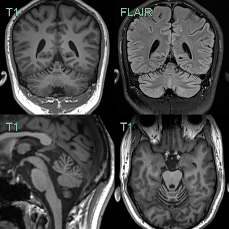
- A 30-year-old male presented with progressive myoclonus and ataxia.
- MRI showed atrophy affecting the pontine tegmentum, paramedian superior cerebellum and possibly the superior vermis.
Treatment
- No disease-modifying therapy exists; management is symptomatic and multidisciplinary:
- Antiseizure medication and treatment of myoclonus in juvenile-onset disease.
- Symptomatic management of chorea, ataxia, and psychiatric features; supportive care and genetic counseling (including implications of paternal anticipation).
- Prognosis is progressive decline over years to decades; earlier onset and longer CAG repeats predict faster progression.
- Imaging role after diagnosis is limited; MRI may be repeated to document progression, exclude comorbid pathology, or evaluate new focal deficits or seizures. Progressive atrophy and expanding white matter signal change are expected and should not be misread as a superimposed acquired leukoencephalopathy.
Differential diagnosis
| Differential diagnosis | Differentiating feature |
|---|---|
| Huntington disease | Prominent caudate/putaminal atrophy with "boxcar" frontal horns; lacks cerebellar/pontine tegmental atrophy and diffuse leukoencephalopathy; HTT CAG expansion |
| Spinocerebellar ataxias (SCA1, 2, 3, 7) | Olivopontocerebellar or pure cerebellar atrophy involving the pontine base; diffuse cerebral leukoencephalopathy is not a typical feature |
| Multiple system atrophy, cerebellar type (MSA-C) | Sporadic, older adults; pontine base atrophy with "hot cross bun" sign, middle cerebellar peduncle T2 hyperintensity, putaminal changes; no heritable pattern |
| CADASIL | White matter disease with characteristic anterior temporal pole and external capsule involvement, lacunes and microbleeds; no cerebellar/tegmental atrophy pattern; NOTCH3 mutation |
| Mitochondrial disease (e.g., MELAS) | Stroke-like cortical lesions crossing vascular territories, lactate on MRS, basal ganglia calcification; episodic clinical course |
| Adult-onset leukodystrophies (e.g., ALSP/CSF1R, adrenoleukodystrophy) | Leukoencephalopathy-predominant with distinct distributions (frontal-predominant with corpus callosum thinning in ALSP; parieto-occipital enhancing margins in ALD); cerebellar/tegmental atrophy not the dominant feature |
| Other progressive myoclonic epilepsies (Lafora disease, Unverricht–Lundborg) | Juvenile myoclonic epilepsy phenotype but MRI typically normal or shows mild diffuse atrophy without leukoencephalopathy or tegmental atrophy |
| Fragile X–associated tremor/ataxia syndrome (FXTAS) | Older males; middle cerebellar peduncle T2 hyperintensity (MCP sign), splenial involvement; FMR1 premutation |
-
Koide et al. Unstable expansion of CAG repeat in hereditary dentatorubral-pallidoluysian atrophy (DRPLA). 1994. Nature genetics - Open in new tab. ↩