Diffuse Axonal Injury (DAI)

- 20-year-old male suffered a severe head injury following a skiing accident.
- T2-weighted imaging showed mature contusional damage at the vertex.
- SWI shows microhemorrhages at the grey-white matter interface, corpus callosum and brainstem.
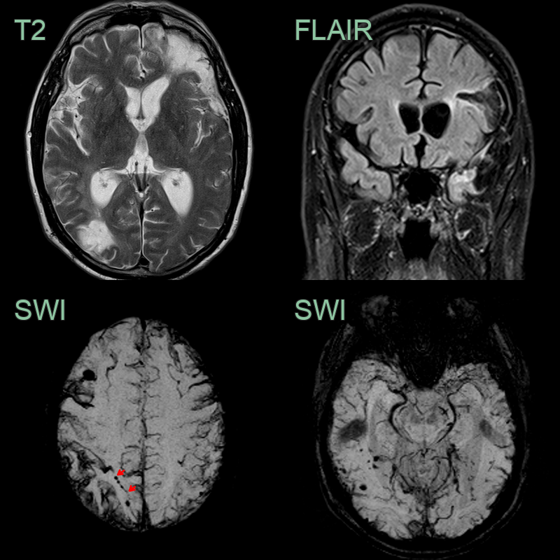
- A 50-year-old patient suffered a severe head injury following a road traffic accident 3 years prior to presentation within worsening cognition.
- MRI showed old parenchymal contusions in the left frontal and temporal lobes alongside an old contre-coup injury in the right occipital lobe.
- SWI showed extensive superficial cortical siderosis secondary to traumatic subarachnoid haemorrhage.
- There were many cortical or immediately subcortical microhaemorrhages. Some of the microhaemorrhages (e.g., in the right superior parietal lobe) were arranged linearly (red arrows).