Diffuse midline glioma
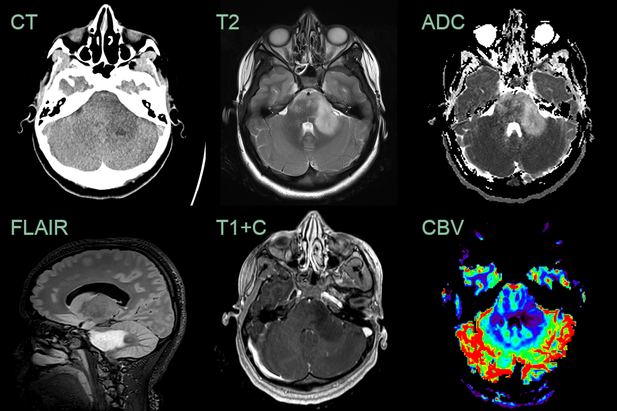
- 20-year-old patient presetned with headaches, blurred vision, nausea and vomitting.
- MRI showed a diffuse T2-hyperintense lesion centred in a mildly expanded cerebellar peduncle.
- Lower ADC values, potentially representing areas of higher celluarity, corresponded to a region of mildly increased rCBV (1.4 relative to the contralateral side).
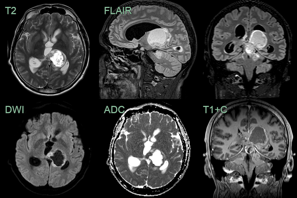
- 20-year-old patient presented with acute onset headache.
- MRI showed a acute obstructive hydrocephalus secondary to a solid-cystic lesion centred on the left thalamus.
- Low ADC values within the solid and enhancing component of the tumor indicated hyperceullarity.
- Biopsy revealed a H3 K27M-mutant diffuse midline glioma.
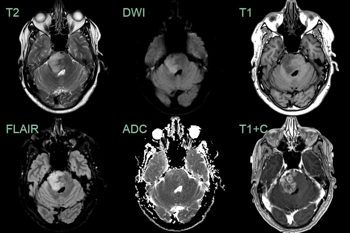
- A 55-year-old patient presented with headache, nausea and vomitting.
- MRI showed an enhancing lesion in the right side of the pons with slightly reduced ADC values.
- Given an extensive travel history the imaging differential included both neoplasta and infection/inflammation.
- Biopsy revealed an H3 27M-mutant diffuse midline glioma.