Enlarged Perivascular Spaces
Summary
- Enlarged perivascular spaces (EPVS) are fluid-filled cavities surrounding small blood vessels in the brain
- They are commonly seen on neuroimaging, particularly in older individuals
- EPVS are generally considered a benign finding but may be associated with various neurological conditions
Pathophysiology
- EPVS, also known as Virchow-Robin spaces, are extensions of the subarachnoid space
- They contain interstitial fluid and follow the course of penetrating arteries and veins
- Enlargement occurs due to:
- Increased fluid accumulation
- Impaired drainage of interstitial fluid
- Alterations in blood-brain barrier permeability
- Associated with:
- Normal ageing
- Cerebral small vessel disease
- Inflammation
- Hypertension
Demographics
- Prevalence increases with age
- More common in:
- Elderly individuals
- Patients with hypertension
- Those with cerebral small vessel disease
- No significant gender predilection reported
Diagnosis
- Often an incidental finding on neuroimaging
- Clinical presentation:
- Usually asymptomatic
- Rarely, may cause mass effect leading to focal neurological deficits
- Differential diagnosis:
- Lacunar infarcts
- Cystic periventricular leukomalacia
- Multiple sclerosis lesions
Imaging
- MRI is the modality of choice for detecting EPVS
- Characteristics on MRI:
- T2-weighted and FLAIR sequences: Hyperintense signal
- T1-weighted sequences: Hypointense signal
- Follow the course of penetrating vessels
- No enhancement with contrast
- Common locations:
- Basal ganglia
- Centrum semiovale
- Midbrain
- Grading systems:
- Based on number and size of EPVS
- E.g., Wardlaw scale: 0 (none) to 4 (severe)
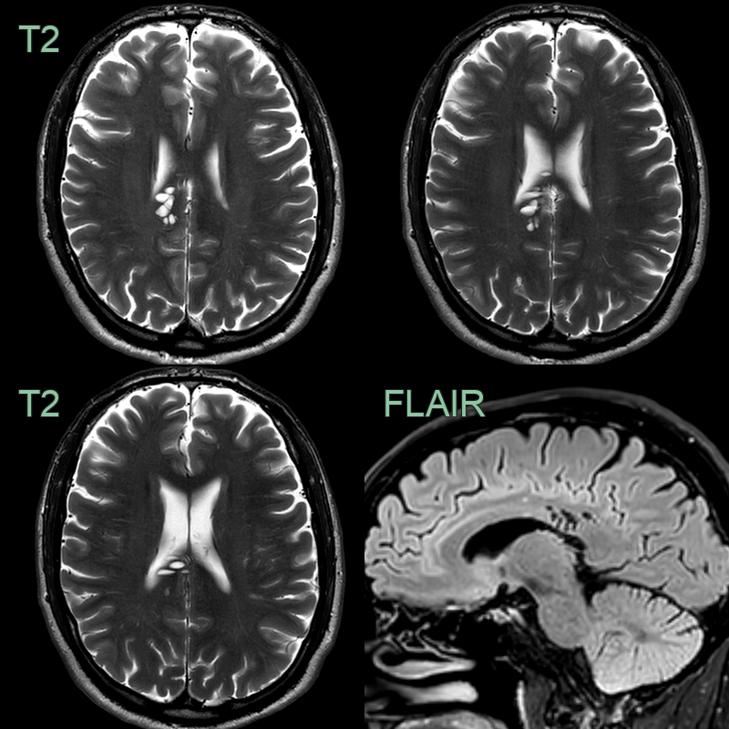
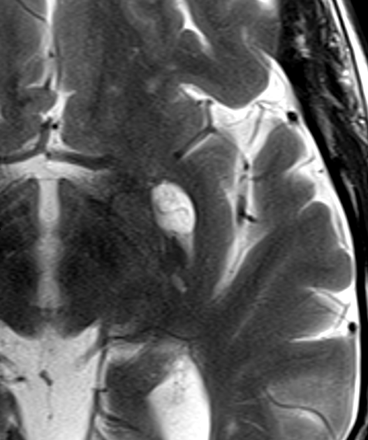
- A cluster of T2-hyperintense lesions centred on the right cingulum fully suppressed on FLAIR and had no surrounding parenchymal signal change.
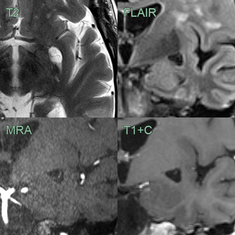
- There is an enlarged perivascular space (T2-hyperintense and fully suppressing on FLAIR) in a typical location; in the subganglionic region.
- Both T2-weighted, time-of-flight angiography and post-gadolinium T1-weighted imaging showed the vessel traversing the perivascular space.
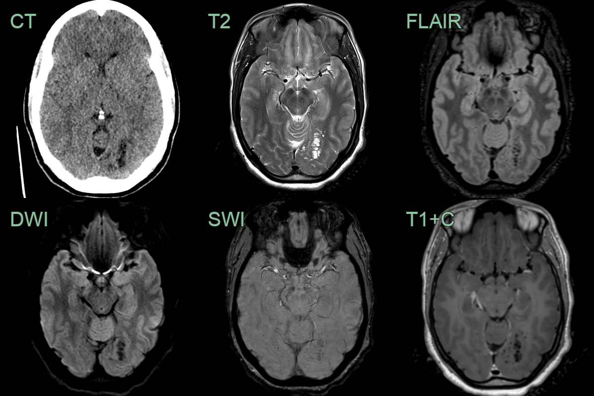
- Incidental finding of a clustered subcortical CSF signal without mass effect was consistent with enlarged perivascular spaces.
- With no FLAIR hyperintensity, an MVNT was not likely.
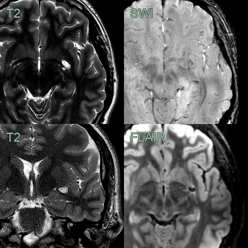
- An incidental enlarged perivascular space in the left subganglionic region.
- The transiting artery is seens as a flow void on T2-weighted imaging.
Treatment
- No specific treatment required for asymptomatic EPVS
- Management focuses on underlying conditions:
- Blood pressure control
- Management of cerebrovascular risk factors
- In rare cases of symptomatic EPVS:
- Surgical decompression may be considered
- Cerebrospinal fluid diversion procedures
Differential diagnosis
| Differential Diagnosis | Differentiating Feature |
|---|---|
| Lacunar infarcts | Irregular shape, surrounding gliosis on FLAIR |
| Multiple sclerosis lesions | Ovoid shape, periventricular predilection, enhancement |
| Small vessel ischaemic changes | Irregular margins, hyperintense on FLAIR |
| Cystic neoplasms | Mass effect, enhancement, irregular borders |
| Neurocysticercosis | Eccentric scolex, surrounding oedema, enhancement |
| Cryptococcosis | Gelatinous pseudocysts, meningeal enhancement |
| Mucopolysaccharidosis | White matter abnormalities, skeletal dysplasia |
| Arachnoid cysts | Larger size, displacement of adjacent structures |
| Adrenoleukodystrophy | White matter involvement, contrast enhancement |