Focal Cortical Dysplasia (FCD)

- 30-year-old patient with frontal lobe seizures.
- MRI showed a subtle region of cortical thickening at the depth of the inferior frontal sulcus.
- A hyperintense tail extending towards the lateral ventricle indicated a Type II focal cortical dysplasia.
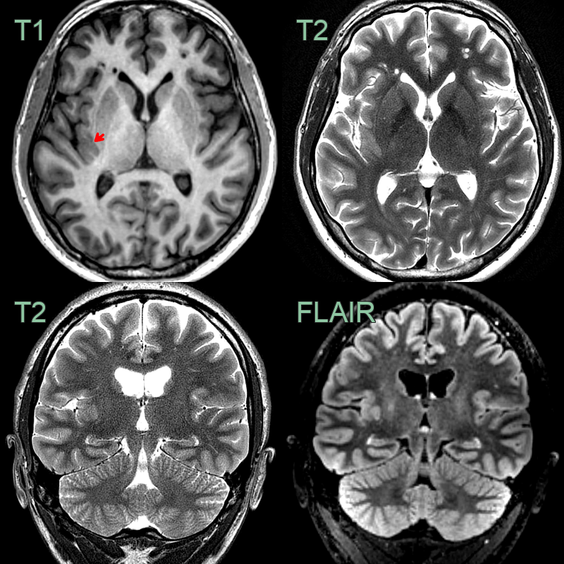
- 25-year-old male presenting with left hand sensory aura.
- MRI showed thickened cortex of the long gyri of the right insula (red arrow) consistent with a focal cortical displasia.
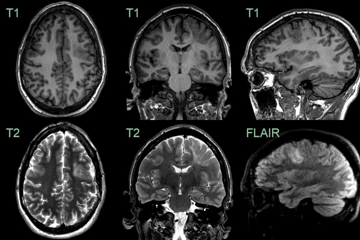
- A 30-year-old patient with epilepsy since childhood.
- MRI showed cortical thickening and subcortical T2-hyperintensity that was consistent with a FCD.