Free Floating Thrombus
Summary
- Free floating thrombus (FFT) is a rare but potentially life-threatening condition characterised by a mobile thrombus in the cardiovascular system, typically attached to a vessel wall by a thin stalk
- FFTs are associated with high risk of embolization and subsequent ischaemic events
- Prompt diagnosis and treatment are crucial to prevent serious complications
Pathophysiology
- FFTs form when a thrombus develops and remains partially attached to the vessel wall
- Common locations include:
- Left atrium (most frequent)
- Aorta
- Carotid arteries
- Deep veins of the lower extremities
- Risk factors:
- Hypercoagulable states
- Atrial fibrillation
- Atherosclerosis
- Trauma
- Malignancy
Demographics
- Incidence is not well-established due to rarity of the condition
- More common in elderly patients
- Higher prevalence in patients with:
- Cardiovascular disease
- History of thromboembolism
- Atrial fibrillation
Diagnosis
- Often an incidental finding on imaging studies
- Clinical presentation may include:
- Symptoms of embolization (e.g., stroke, limb ischaemia)
- Asymptomatic in some cases
- Diagnostic criteria:
- Mobile thrombus
- Thin stalk attachment to vessel wall
- Independent motion from surrounding structures
Imaging
- Echocardiography:
- Transthoracic echocardiography (TTE): initial screening tool
- Transesophageal echocardiography (TEE): gold standard for cardiac FFTs
- CT angiography:
- High sensitivity for detecting FFTs in large vessels
- Allows for evaluation of surrounding anatomy
- MRI:
- Useful for characterising thrombus composition
- Can differentiate between thrombus and tumour
- Doppler ultrasound:
- Valuable for detecting FFTs in peripheral vessels
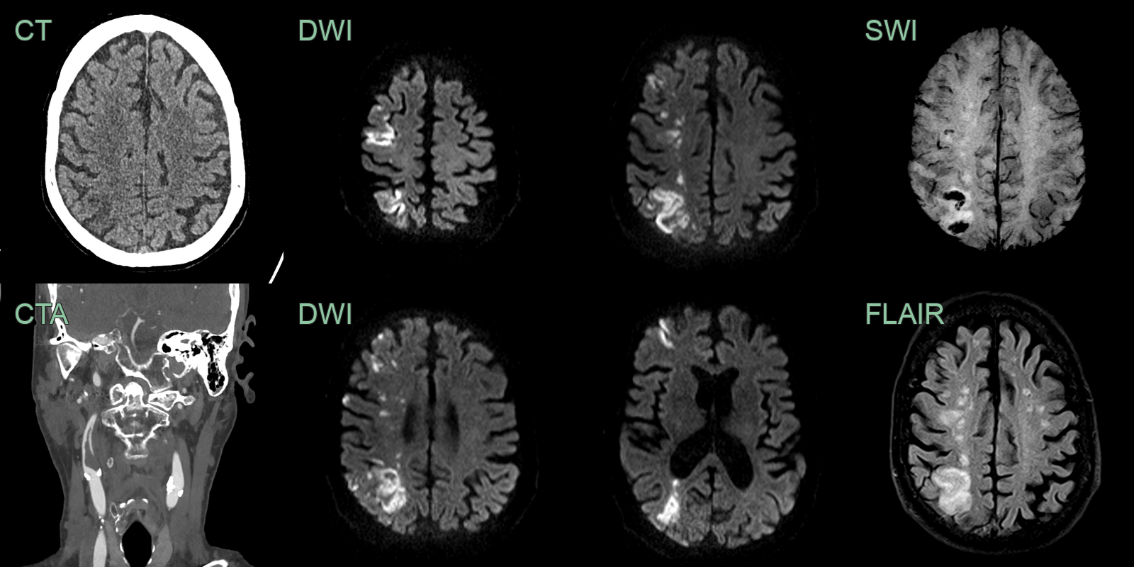
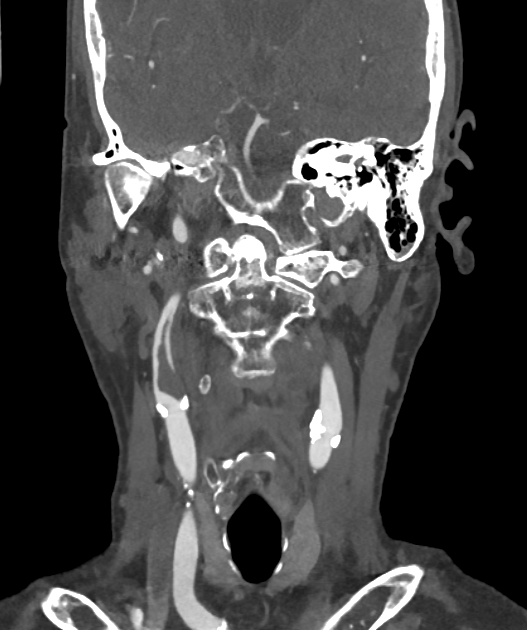
- 60-year-old patient with left sided weakness and inattention.
- CTA showed acute thrombus arising from plaque in the right ICA.
- The filling defect extending distally within the lumen of the vessel was consistent with free floating thrombus.
Treatment
- Anticoagulation:
- First-line treatment for most FFTs
- Heparin followed by oral anticoagulants (e.g., warfarin, direct oral anticoagulants)
- Thrombolysis:
- Consider in selected cases
- Risk of thrombus fragmentation and embolization
- Surgical intervention:
- Indicated for large FFTs or those refractory to medical management
- Thrombectomy or embolectomy
- Endovascular approaches:
- Catheter-directed thrombolysis
- Mechanical thrombectomy devices
- Follow-up imaging:
- Regular monitoring to assess thrombus resolution and detect recurrence
Differential diagnosis
| Differential Diagnosis | Differentiating Feature |
|---|---|
| Aortic dissection | Linear intimal flap on imaging |
| Aortic atheroma | Calcification and plaque on vessel wall |
| Cardiac myxoma | Attachment to interatrial septum |
| Vegetation | Associated with valvular lesions |
| Tumour embolus | History of malignancy |
| Arteritis | Vessel wall thickening and inflammation |
| Intramural haematoma | Crescentic thickening of vessel wall |
| Artefact on imaging | Disappears with different imaging views |
| Papillary fibroelastoma | Typically attached to cardiac valves |
| Aortic aneurysm with thrombus | Dilated aortic segment |