Mogad
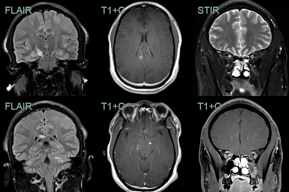
- A 30-year-old patient presented with ataxia and optic neuritis.
- MRI showed multiple enhancing lesions in the cerebellum, cerebral peduncles, corpus callosum and intraorbital right optic nerve.
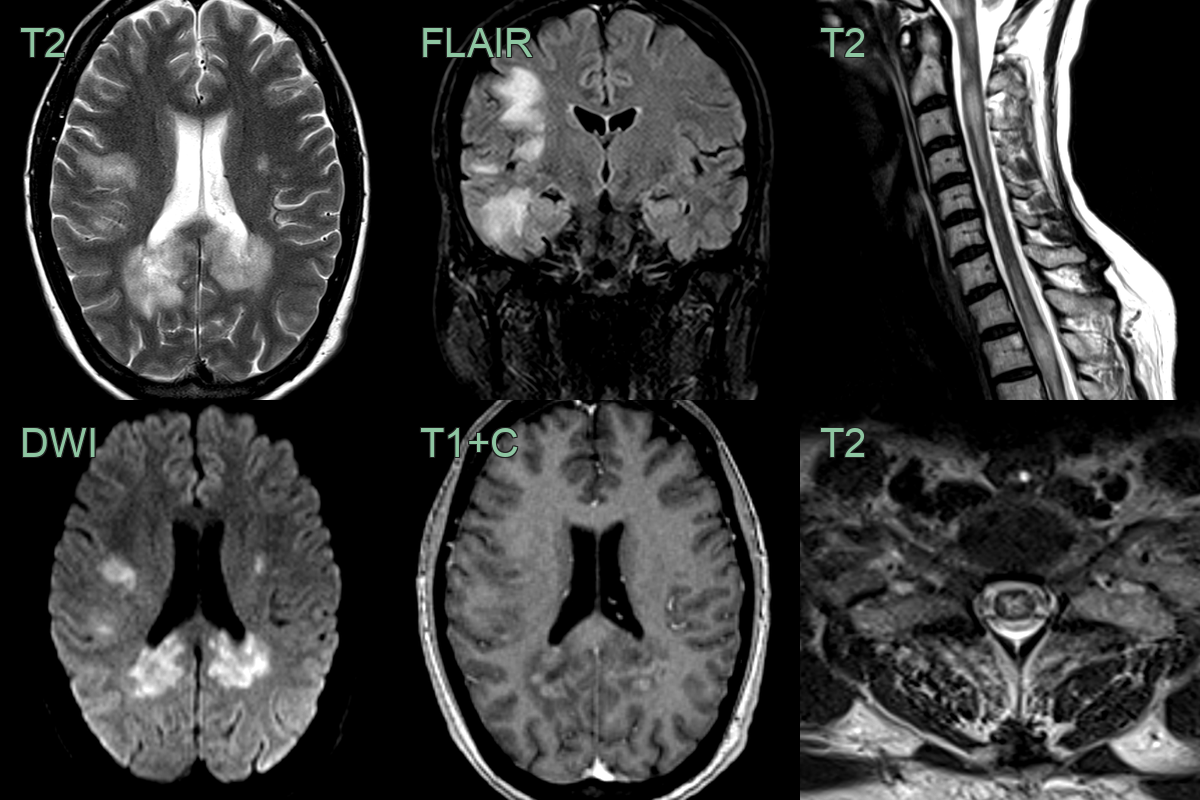
- A 40 year old presented with confusion and upper and lower limb weakness and sensory disturbance.
- MRI showed extensive white matter lesions within both cerebral hemispheres associated with diffusion restriction and peripheral enhancement.
- In the cord, there were multiple swollen mainly central short segment cord lesions.