Neurofibromatosis Type 1
Summary
- Autosomal dominant genetic disorder characterised by café-au-lait spots, neurofibromas, and Lisch nodules
- Caused by mutations in the NF1 gene, leading to uncontrolled cell growth and tumour formation
- Imaging plays a crucial role in diagnosis, monitoring, and management of associated complications
Pathophysiology
- Mutation in the NF1 gene on chromosome 17q11.2
- NF1 gene encodes neurofibromin, a tumour suppressor protein
- Loss of neurofibromin function leads to:
- Increased Ras signalling
- Uncontrolled cell proliferation
- Formation of benign and malignant tumours
Demographics
- Incidence: 1 in 2,500 to 3,000 live births
- No ethnic or gender predilection
- 50% of cases are familial, 50% are due to de novo mutations
- Complete penetrance with variable expressivity
Diagnosis
- Clinical diagnosis based on NIH diagnostic criteria :
- Six or more café-au-lait macules (>5 mm in prepubertal individuals, >15 mm in postpubertal individuals)
- Two or more neurofibromas or one plexiform neurofibroma
- Axillary or inguinal freckling
- Optic glioma
- Two or more Lisch nodules
- Distinctive osseous lesion (sphenoid dysplasia or tibial pseudarthrosis)
- First-degree relative with NF1
- Genetic testing for NF1 gene mutations
Imaging
- MRI:
- Brain:
- Unidentified bright objects (UBOs) in basal ganglia, thalamus, and cerebellum
- Optic pathway gliomas
- Plexiform neurofibromas
- Spine:
- Dural ectasia
- Spinal neurofibromas
- CT:
- Skeletal abnormalities:
- Sphenoid wing dysplasia
- Tibial pseudarthrosis
- Scoliosis
- Ultrasound:
- Superficial plexiform neurofibromas
- Screening for renal artery stenosis
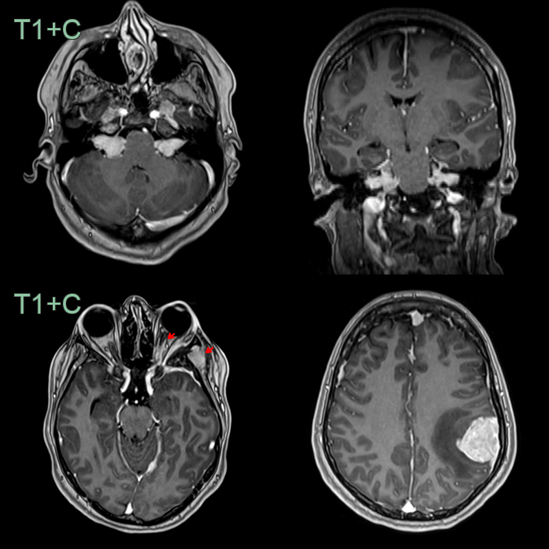
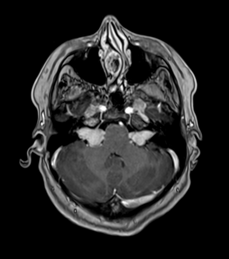
- A 20-year-old patient presented after a seizure.
- MRI showed bilateral vestibular schwannomas and many meningiomas (optic nerve sheath [red arrow], anterior falx and left parietal region).
Treatment
- Multidisciplinary approach
- Regular clinical and imaging follow-up
- Surgical management:
- Symptomatic or rapidly growing neurofibromas
- Optic pathway gliomas causing visual impairment
- Medical management:
- Selumetinib for inoperable plexiform neurofibromas
- Pain management
- Treatment of associated conditions (e.g., hypertension, epilepsy)
- Genetic counselling
- Psychosocial support
Differential diagnosis
| Differential Diagnosis | Distinguishing Feature |
|---|---|
| Legius syndrome | Absence of neurofibromas and optic gliomas |
| McCune-Albright syndrome | Café-au-lait spots with irregular borders ("coast of Maine") |
| Multiple endocrine neoplasia type 2B | Presence of medullary thyroid carcinoma and mucosal neuromas |
| Tuberous sclerosis | Presence of ash-leaf spots and facial angiofibromas |
| Proteus syndrome | Asymmetric overgrowth and vascular malformations |
| Klippel-Trenaunay syndrome | Vascular malformations and limb hypertrophy |
| Bannayan-Riley-Ruvalcaba syndrome | Macrocephaly and intestinal polyposis |
| LEOPARD syndrome | Presence of lentigines and cardiac abnormalities |
| Noonan syndrome | Distinctive facial features and congenital heart defects |
| Mosaic neurofibromatosis | Localised or segmental distribution of neurofibromas |