Olfactory Neuroblastoma
Summary
- Rare malignant neuroectodermal tumour arising from the olfactory epithelium
- Typically presents with unilateral nasal obstruction and epistaxis
- Characteristic imaging findings include a dumbbell-shaped mass in the superior nasal cavity extending into the anterior cranial fossa
Pathophysiology
- Originates from neural crest cells in the olfactory epithelium
- Composed of small round blue cells with neuroendocrine differentiation
- Molecular alterations include:
- IDH2 mutations (82.6% of cases)
- TP53 mutations (20-25% of cases)
- CDKN2A/B deletions (30-50% of cases)
Demographics
- Accounts for 3-6% of intranasal tumours
- Bimodal age distribution:
- First peak: 10-20 years
- Second peak: 50-60 years
- Slight male predominance (1.2:1)
- No known racial predilection
Diagnosis
- Clinical presentation:
- Unilateral nasal obstruction (70%)
- Epistaxis (50%)
- Anosmia (40%)
- Headache (20%)
- Endoscopic examination:
- Polypoid, friable mass in the superior nasal cavity
- Biopsy:
- Essential for definitive diagnosis
- Immunohistochemistry: positive for synaptophysin, chromogranin, and neuron-specific enolase
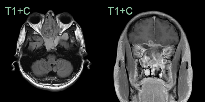
Imaging
- CT findings:
- Homogeneous soft tissue mass in the superior nasal cavity
- Bone erosion of the cribriform plate and lamina papyracea
- Calcifications in 20-25% of cases
- MRI findings:
- T1: isointense to gray matter
- T2: heterogeneous signal intensity
- T1 post-contrast: moderate to intense enhancement
- Characteristic "dumbbell shape" with intracranial and intranasal components
- PET/CT:
- Intense FDG uptake (SUVmax > 10)
- Useful for staging and detecting distant metastases
Treatment
- Multimodal approach:
- Surgery:
- Endoscopic resection for early-stage tumours
- Craniofacial resection for advanced tumours
- Radiation therapy:
- Adjuvant radiotherapy (54-66 Gy) for local control
- Chemotherapy:
- Reserved for advanced or metastatic disease
- Regimens include cisplatin/etoposide or cyclophosphamide/vincristine/doxorubicin
- Prognosis:
- 5-year overall survival: 60-80%
- Factors associated with poor prognosis:
- Advanced Kadish stage
- High-grade histology
- Intracranial extension
- Follow-up:
- Regular imaging (MRI) every 3-6 months for the first 2 years, then annually
- Long-term surveillance due to risk of late recurrence
Differential diagnosis
| Differential Diagnosis | Differentiating Feature |
|---|---|
| Sinonasal carcinoma (SCC/SNUC) | May appear identical on imaging; typically lacks peritumoural cysts; often more aggressive bone destruction |
| Sinonasal melanoma | T1 shortening (hyperintensity) due to melanin content; aggressive bone destruction |
| Lymphoma | Homogeneous soft tissue mass; may appear identical; lacks peritumoural cysts; no calcification |
| Olfactory groove meningioma | Dural attachment with dural tail; hyperostosis; superior extension from above cribriform plate |
| Nasopharyngeal carcinoma | Epicentre more posteriorly located in nasopharynx; cervical nodal metastases |
| Rhabdomyosarcoma | More common in children; hypointense T2 signal; may involve orbit |
| Inverted papilloma | Cerebriform T2 signal pattern; unilateral; focal hyperostosis at site of origin; no intracranial extension |