Ossification of the falx cerebri
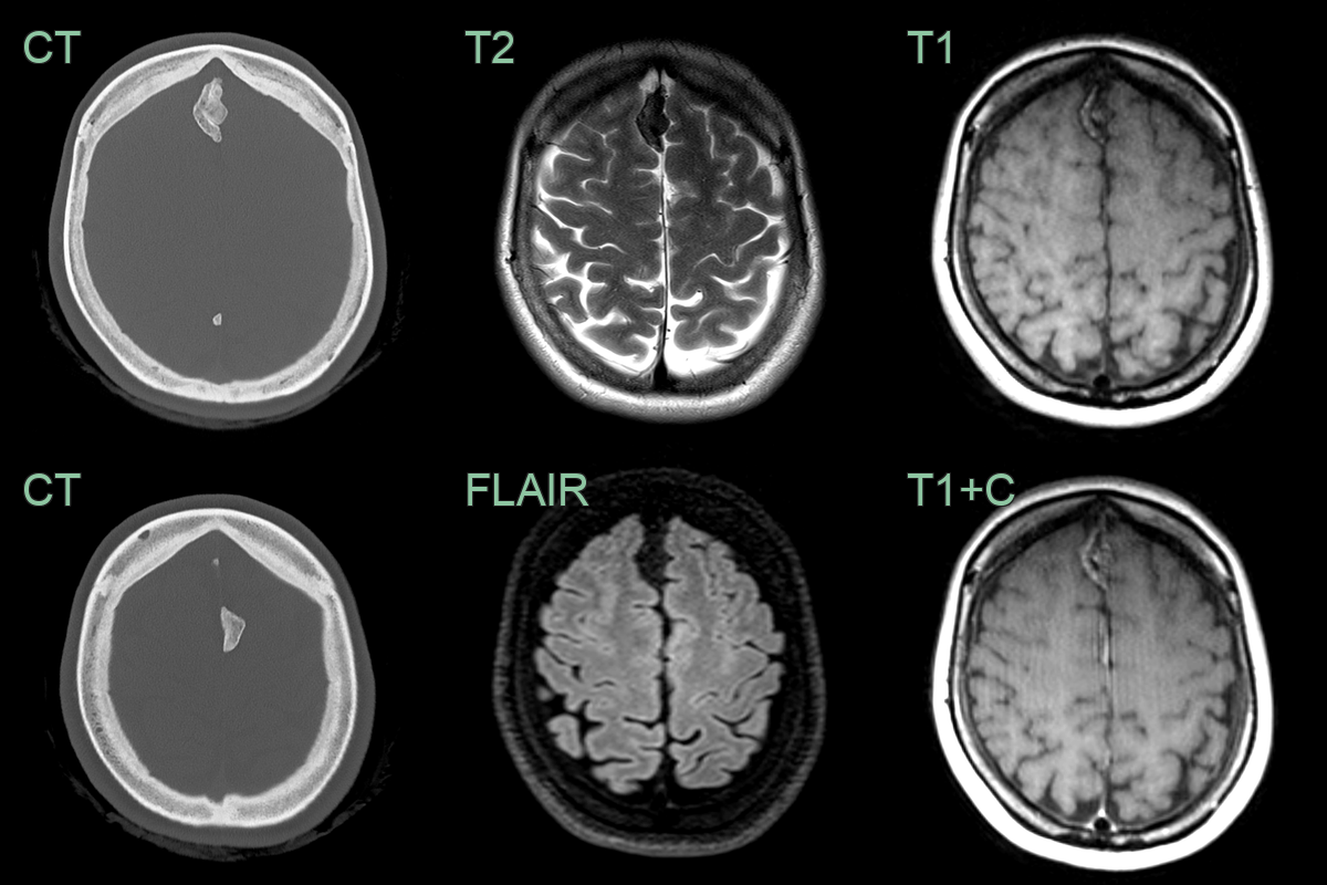
- A 50-year-old patient presented with headache.
- CT showed lobulated calcification of both sides of the falx.
- The T1-hyperintensity and lack of enhancement was consistent with ossification of the falx (rather than a meningioma).