Spinal CSF leak
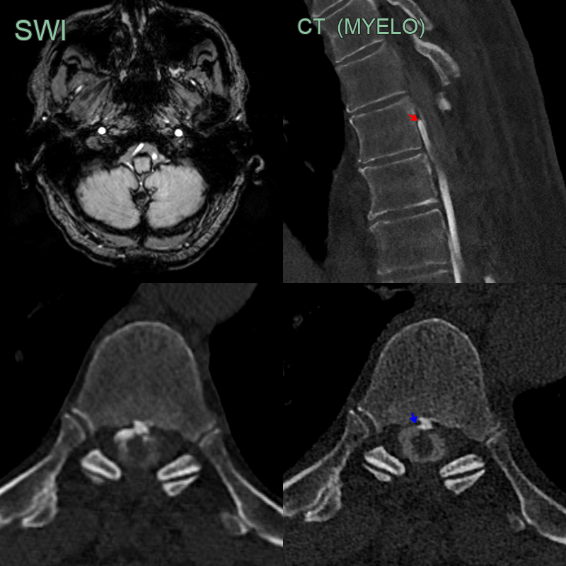
- 70-year-old patient with orthostatic headaches and transient bilateral 6th nerve palsies. The patient had a multi-level thoracic laminectomy for a compressive arachnoid cyst many years prior.
- CT myelography showed a rapidly filling small ventral epidural leak (red arrow).
- More apparent on later phase imaging, the ventral leak was associated with a small osteophyte (blue arrow).
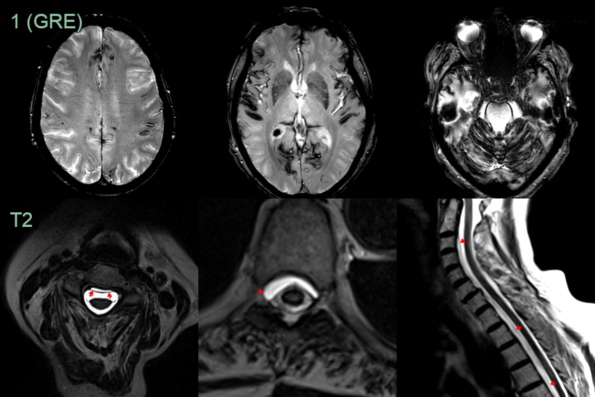
- A 70-year-old patient presented with tinnitus and dizziness.
- MRI showed extensive superficial siderosis above and below the tentorium.
- While the patient had no headache, given the distribution of siderosis, a CSF leak was suspected.
- MRI of the spine showed a longitudinally extensive ventral epidural collection, indicating a CSF leak.