Case of the Month: November 2025
Case history
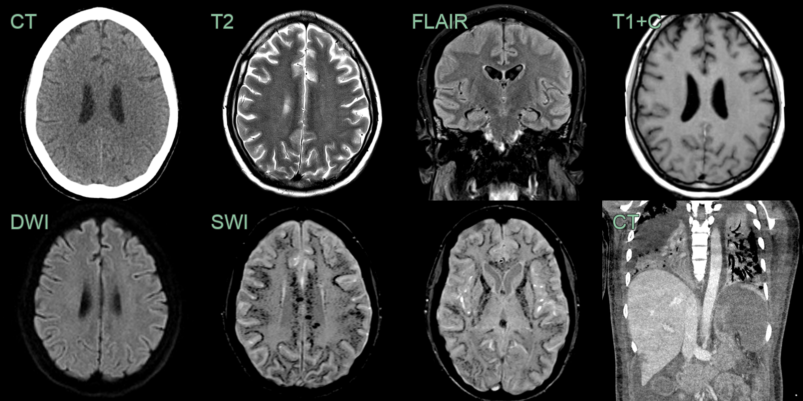
- 40-year-old patient became obtunded 1 month after returning from a region endemic for malaria.
Click to reveal diagnosis
- The brain parenchyma appeared normal on T2, FLAIR, and DWI — there was no oedema or ischaemic change.
- SWI showed extensive juxtacortical and deep white matter foci of susceptibility artefact, representing microhaemorrhages and/or microthrombi.
-
Chest imaging showed bilateral lung consolidation, splenomegaly with infarction, and small regions of hepatic infarcts.